
Ureterocalicostomy
Ureterocalicostomy is a reconstructive kidney surgery that connects the ureter directly to a lower part of the kidney, bypassing a scarred or blocked section so urine can drain freely again. It is used mainly when a standard repair has failed or isn't possible.

Briefly about this treatment
Indications
Complex or repeat ureteropelvic junction (UPJ) obstruction, scarring after a failed pyeloplasty, a renal pelvis that sits inside the kidney, or a proximal ureteral stricture that can't be repaired in the standard way.
Risks
Urine leakage at the new connection, infection, bleeding, and re-narrowing of the join. Risks are minimised by careful patient selection and surgeons experienced in ureteral reconstruction.
Pre-procedure
Blood and urine tests, imaging (CT urogram or retrograde pyelogram, plus a kidney scan to check function), anaesthesia consultation, fasting per hospital protocol.
Follow-up
Imaging and clinic visits in the first weeks and months, stent removal typically after about 5 weeks to 3 months, then long-term monitoring of drainage and kidney function; the exact schedule is set by your surgeon.
What is Ureterocalicostomy? An expert explains
The name breaks down literally: uretero- (ureter) + calico- (calyx, the cup-shaped part of the kidney that collects urine before it drains) + -stomy (a surgical connection). So the procedure connects the ureter to a kidney calyx instead of to the renal pelvis.
Surgeons reach for it in specific situations, most often:
- UPJ obstruction where the renal pelvis sits mostly inside the kidney (an „intrarenal pelvis”), making the usual repair technically difficult.
- A previous pyeloplasty that failed, leaving scar tissue around the UPJ.
- A proximal ureteral stricture after stone treatment (such as ureteroscopy or percutaneous stone removal) or after a failed endoscopic incision of the UPJ.
Signs it may be time for Ureterocalicostomy
No symptom on its own means you need this surgery. It is considered when urine drainage from one kidney stays blocked despite earlier treatment, and your urologist confirms it with imaging and a kidney-function scan. These are the things people commonly notice:
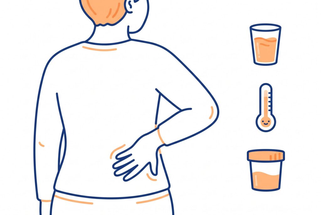
- Flank or back pain on one side that keeps returning, sometimes worse after drinking a lot of fluid or alcohol. Pain that comes back after a previous kidney or ureter operation is a key reason to ask about other options.
- Repeated urinary tract infections or fever with flank pain, because urine that can’t drain freely is more likely to become infected.
- Blood in the urine, which may look pink, red, or tea-coloured.
- Nausea or vomiting during pain episodes, which can happen when the kidney is stretched by backed-up urine.
Conditions linked to ureterocalicostomy
These are the conditions where your urologist may discuss this surgery. Each links to its own page.
- Ureteropelvic Junction Obstruction — The blockage where the kidney meets the ureter; ureterocalicostomy is mainly used when this obstruction is complex, scarred, or has come back after a standard repair.
- Hydronephrosis — Swelling of the kidney from backed-up urine, which is the main thing the surgery aims to relieve.
- Horseshoe Kidney — When the kidneys are fused at the lower poles, a UPJ blockage can be harder to repair the usual way; children and adults with fusion or rotation anomalies are among those treated with this surgery .
- Obstructive Uropathy — The wider group of urine-flow blockages that can harm kidney function, which this procedure treats at the top of the ureter.
- Nephrolithiasis (Kidney Stones) — Scarring or narrowing of the upper ureter after stone removal procedures is one of the situations that leads to this surgery.

When to consult a specialist
Flank pain and repeat infections are common, and they very often have a treatable cause. A simple blood test (creatinine and eGFR) and a urine test can show how well your kidneys are working, and a painless ultrasound can show whether urine is draining. If you’ve already had kidney, ureter, or stone surgery and your symptoms have come back, that’s a good reason to ask for a second look — not a reason to worry. When you’re ready, a urologist experienced in kidney and ureter reconstruction can walk you through your options at your own pace.
How a session works
Ureterocalicostomy is a single operation rather than a series of sessions. It can be done through an open incision, laparoscopically (small incisions with a camera), or with robotic assistance, depending on your anatomy, your surgeon’s experience, and any previous surgery.
- Before surgery — imaging (CT urogram and/or a retrograde pyelogram) confirms exactly where the blockage is and maps the kidney’s anatomy.
- Anesthesia and access — under general anesthesia, the surgeon reaches the kidney through a flank or abdominal incision (open) or several small ports (laparoscopic/robotic).
- Exposing the kidney — the retroperitoneum, proximal ureter, and lower part of the kidney (including the blood-vessel-rich hilum) are carefully dissected free.
- Removing the diseased segment — a small, targeted portion of scarred lower-pole kidney tissue is removed (a modest partial nephrectomy) to reach a healthy, dependent calyx.
- The anastomosis — the ureter is opened at an angle (spatulated) and sutured directly onto the calyx, creating a new, wide drainage channel that bypasses the damaged UPJ.
- Stents and drains — a temporary Double-J ureteral stent, and sometimes a nephrostomy tube or abdominal drain, is placed to protect the new connection while it heals.
- Closing — incisions are closed and you’re moved to recovery.
Benefits
The main reason for choosing this surgery is to keep a kidney that might otherwise be lost. It gives the kidney a new, unobstructed way to drain when the usual repair isn’t possible.
- Kidney preservation. It is described in the surgical literature as a salvage or last-resort option for protecting a working kidney that scarring has put at risk, as an alternative to removing it.
- Better drainage and function. In one adult series, the treated kidney’s share of total function rose from an average of 54.6% before surgery to 60.1% after, and blockage was relieved in every patient. That series was small, so individual results vary.
- Symptom relief. In a series of 22 adults, 16 (73%) were cured or improved after about two years of follow-up.
- Works when other repairs can’t. It can be the best option for complex UPJ obstruction with an intrarenal pelvis, or where the kidney’s drainage space is more swollen than the pelvis.
- Keyhole and robotic options. In small comparisons, the laparoscopic approach had a shorter operating time and less blood loss than open surgery, with no clear difference in hospital stay.
Risks & side effects
Ureterocalicostomy has risks and trade-offs to discuss with your care team. The information below comes from published surgical studies and this treatment’s data.
Urine leakage, infection, bleeding, and re-narrowing of the new connection. Risks are minimised by careful patient selection and surgeons experienced in ureteral reconstruction.
- Urine leakage where the ureter joins the kidney, which usually settles with drainage while the join heals.
- Infection of the wound, the urine, or the tissue around the kidney.
- Bleeding, as with any kidney operation, since the surgery involves a small part of the kidney tissue.
- Re-narrowing of the new connection, which is the main long-term risk and can mean further treatment.
- Stent discomfort while the temporary tube is in place: some people feel urgency, a need to pass urine more often, or mild flank ache, which usually eases once it is removed.
- General surgery and anesthesia risks, such as blood clots, chest problems, and reactions to medication.
Who it may not suit: People whose kidney has too little working function left to be worth preserving, people with an active uncontrolled infection, and people for whom a standard or repeat pyeloplasty is still possible. These are general cautions, not a personal verdict. A qualified clinician must assess your full medical picture before recommending or delaying treatment.

What happens during & after
Operating time varies with the approach and how complex the anatomy is.
Immediately afterward, you’ll wake up with a urinary catheter and, usually, the ureteral stent already in place. Pain is managed with medication, and imaging or lab checks confirm the kidney is stable before you’re discharged.
Possible complications
Most people do well, but a small number run into problems that need extra treatment. These figures come from small surgical case series (a few dozen patients each, not large trials), so ask your own team about outcomes specific to their experience and your case.
- Re-narrowing or failure of the new join. In a series of 22 adults followed for about 27 months, the operation failed in 4 and left 2 unchanged. A further narrowing can sometimes be treated again, and occasionally the kidney can’t be saved and needs to be removed — one case report describes narrowing with a pus-filled, blocked kidney that required removal.
- Prolonged urine leak or infection around the kidney. In a series of 21 children, 19 had excellent results and 2 had complications: a prolonged leak at the join in one child, and a fungal (Candida) abscess around the kidney in another.
- Ongoing discomfort despite better drainage. In a small laparoscopic series, one patient still had flank discomfort even though scans showed improved drainage, and chose to have the kidney removed at 6 months.
- Few problems in some series. In 11 adults treated with open surgery, none had a significant perioperative complication.
If you notice fever, worsening flank pain, or fluid leaking from the wound or drain site, contact your surgical team promptly.
New or severe symptoms require prompt medical advice. Do not wait for a routine appointment if you feel seriously unwell.
Recovery
Most people stay in hospital for several days, then go home with a temporary stent that stays in place for several weeks to a few months while the new connection heals.
- Hospital stay. Open-surgery series report about 4–7 days, averaging around 5; small laparoscopic series report stays as short as 2 days. In one robotic case, the abdominal drain came out on day one. Your own stay depends on the approach and how you recover.
- Stent removal. The stent typically comes out after about 5–8 weeks in small series, and one protocol kept it for 2–3 months. Removal is usually a quick outpatient procedure using a thin camera passed through the urethra (cystoscopy).
- Follow-up scans. Your team will check the new connection with imaging — an ultrasound, a kidney scan with a diuretic (renography), or a CT urogram — for example, a CT urogram at 3 months in one robotic case. In one adult series, follow-up ran from 5 to 32 months.
- Activity and return to normal life. Follow your surgeon’s instructions on lifting, driving, and work; these are set individually [needs source: individualized activity guidance].
- Long-term outlook. Published results are generally good, including in previously failed cases, but a minority of people need further treatment for re-narrowing (see section 11). Regular check-ups let any problem be caught early.
This is general information, not a personal recovery plan. Follow the instructions your own surgical team gives you.
Clinics offering Ureterocalicostomy
Verified kidney centres linked to this treatment in the KidneyGlobal directory.



Most Common Questions
Is ureterocalicostomy a common surgery?
No. It is an infrequent, specialized operation, generally kept for complex or repeat cases of UPJ obstruction rather than used as a first repair.
How is it different from pyeloplasty?
In a pyeloplasty, the surgeon rebuilds the kidney's renal pelvis and reconnects the ureter to it. In ureterocalicostomy, the pelvis is bypassed and the ureter is joined directly to a kidney calyx. It is usually reserved for cases where a standard pyeloplasty has failed or isn't workable.
Will I lose kidney function after the surgery?
The aim is to protect the function you have. In small published series, drainage and function improved for many people, but results vary and a few people need further treatment.
Will I need a permanent stent or tube?
Usually not. A temporary ureteral stent, and sometimes a short-term drain or nephrostomy tube, protects the new connection while it heals and is then removed.
Can it be done with keyhole or robotic surgery?
Yes. Laparoscopic and robot-assisted versions have been described, although most people have historically needed open surgery, and the approach depends on your anatomy and your surgeon's experience.
Which doctor performs this operation?
A urologist, usually one with extra training in reconstructive, pediatric, or robotic urology — not a nephrologist. Because the procedure is rare, ask any surgeon how many they have done before you decide.
Sources
- Ureterocalicostomy for treatment of complex cases of ureteropelvic junction obstruction in adultsPubMed, U.S. National Library of Medicine
- Ureterocalicostomy: A contemporary experienceElsevier (ScienceDirect archive; journal Urology)
- Laparoscopic Ureterocalicostomy: Initial ExperienceJournal of Urology, via ScienceDirect (Elsevier)
- Ureterocalicostomy for Renal Salvage (pediatric series)Journal of Urology, via ScienceDirect (Elsevier)
- Ureterocalicostomy for complex upper ureteral stricture: a narrative review of the current literaturePubMed Central, U.S. National Library of Medicine
- Robotic-assisted Laparoscopic Ureterocalicostomy (RALUC): How we do itPubMed Central, U.S. National Library of Medicine
- Ureterocalycostomy — final resort in the management of secondary pelvi-ureteric junction obstruction: our experiencePubMed Central, U.S. National Library of Medicine
- Ureterocalicostomy: a bailout package for kidney preservation in secondary pelviureteric junction obstructionResearchGate (hosted paper)
- AB008. Robotic ureterocalycostomy for complex pelviureteric junction obstructionAME Publishing Company
- Efficacy of ureterocalicostomy in treating secondary long-segment ureteral stricture (stent-duration protocol)e-Century Publishing Corporation
- Ureteropelvic Junction Obstruction: Treatment & ManagementMedscape (eMedicine)
- Ureteropelvic Junction Obstruction (Adult)UCSF Department of Urology
- HCPCS 50750 — Ureterocalycostomy negotiated rates (July 2026)Tynbil, LLC