

Academic Medical Center
Comprehensive Nephrology & Transplant
Joint CommissionMagnet RecognitionUNOS Transplant Center
✓ 24/7 ER✓ ICU✓ Insurance
An MRI urogram is a painless, radiation-free scan that gives your care team detailed images of your kidneys, ureters and bladder — used to find the cause of symptoms like blood in the urine, flank pain, or a suspected blockage.
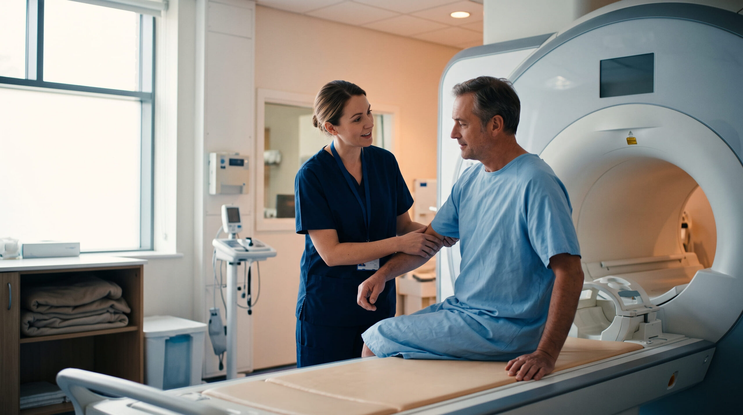
Unexplained blood in the urine, ongoing flank or lower-back pain suggesting a blocked or swollen kidney, recurrent urinary tract infections, a suspected structural problem in the urinary tract, or the need for repeat imaging without radiation exposure.
Rare reactions to gadolinium contrast, discomfort from the enclosed scanner, and limited suitability for certain implants or pregnancy. Risks are minimised through careful pre-scan screening and experienced radiology teams.
Screening for MRI-incompatible implants and pregnancy, removal of metal objects and jewelry, a recent kidney-function blood test if contrast is planned, and — depending on the protocol — fasting or extra fluids beforehand.
No fixed follow-up visits are needed for the scan itself. A radiologist sends a report to your referring doctor, usually within a few days, who then discusses results and any next steps with you.
MRI urogram uses a strong magnetic field and radio waves — not radiation — together with a computer to build detailed images of your urinary tract: the kidneys, the ureters (the tubes that carry urine to your bladder), and the bladder itself. There are two main types your radiologist may use, sometimes together: a static-fluid scan, which uses urine already in your urinary tract as a natural contrast and needs no injection, and an excretory scan, where a gadolinium-based contrast dye is injected so the radiologist can watch how your kidneys filter and drain it over time.
Compared with a CT urogram, an MRI urogram doesn’t use ionizing radiation, which is why it’s often preferred for children, pregnant patients, people needing repeat scans, and those with reduced kidney function or an iodine-contrast allergy. The trade-off: it’s less sensitive than CT for picking up small urinary stones.
You might notice:
These are the conditions linked to this procedure in the directory. Your clinician decides whether this scan is appropriate using your symptoms, examination and test results.
Remember: These entries are not a diagnosis or a treatment recommendation. Discuss your symptoms and test results with a qualified clinician.

If you’ve noticed blood in your urine, ongoing flank pain, or repeated infections, it’s worth getting checked — these are common and often manageable once the cause is found, so try not to jump to the worst conclusion. A simple blood or urine test is often the first step, and your doctor will tell you if imaging like this is needed. You can consult a specialist directly through our find a specialist tool to get a clear answer sooner rather than later.
Check-in and screening. Staff confirm you have no MRI-incompatible implants (pacemakers, certain aneurysm clips, some cochlear implants) and check whether you’re pregnant or could be.
Preparation. You’ll usually remove metal objects, jewelry and anything with a magnetic strip, and change into a gown. Some protocols ask you to avoid solid food for a few hours and drink a specified amount of clear liquid beforehand.
On the table. You lie on a movable exam table, a coil may be placed near the area being studied, and you’ll get earplugs since the scanner is noisy.
Inside the scanner. The table slides into the MRI tunnel. You’ll get breathing instructions at points during the scan to improve image quality.
Contrast (if used). A small IV line delivers gadolinium-based contrast partway through; you may feel a brief cool sensation.
Finishing up. The table slides out, the IV (if placed) is removed, and — unless you were sedated — you can typically get dressed and leave right away.
This scan has risks and trade-offs to discuss with your care team. The information below comes from this procedure’s data. Gadolinium contrast reactions are uncommon and usually mild; certain implants or pacemakers can make the scanner unsafe; and the enclosed tunnel can be uncomfortable for people with claustrophobia. Risks are minimised through careful pre-scan screening and experienced radiology teams.
Who it may not suit: people with certain pacemakers, non-titanium aneurysm clips, or other MRI-incompatible implants; people with significantly reduced kidney function, where contrast needs extra caution; and pregnant patients, where contrast is generally avoided. These are general cautions, not a personal verdict — a qualified clinician must assess your full medical picture before recommending or delaying this scan.

During the scan, the main thing asked of you is to stay still and follow breathing cues. The machine is loud — expect knocking and buzzing sounds, which is normal. If you feel anxious in enclosed spaces, tell the radiology team beforehand; many centers offer wide-bore scanners, a mirror to see out, music, or a mild sedative.
Afterward, if you didn’t have sedation, you can eat, drink and drive as normal right away. If contrast was used, drinking extra water can help your kidneys clear it, unless your doctor advises otherwise. A radiologist reviews the images and sends a report to the doctor who ordered your scan, usually within days.
New or severe symptoms require prompt medical advice. Do not wait for a routine appointment if you feel seriously unwell.
There’s no real “recovery” from an MRI urogram — it isn’t invasive, and most people resume normal activities immediately. Watch for any late reaction at the IV site, such as redness or swelling, or signs of an allergic reaction, and contact the clinic if these occur. Your doctor will discuss what the images show and any next steps, which may include further tests, a specialist referral, or starting treatment for an underlying condition.
Verified kidney centres linked to this treatment in the KidneyGlobal directory.



No. It uses a magnetic field and radio waves, not X-rays.
No — both image the urinary tract, but CT has wider availability and shorter exam times, while MRI gives superior soft-tissue detail and avoids radiation.
Not always. A static-fluid scan needs no contrast; an excretory scan uses an IV gadolinium-based agent, decided based on your kidney function and what your radiologist needs to see.
No. You may feel brief discomfort from lying still, the noise, or a cool sensation if contrast is injected, but the scan itself isn’t painful.
Often yes for the non-contrast version. Whether you can safely have contrast depends on your current kidney function, which your care team checks first.
A radiologist reviews the images and sends a report to your referring doctor, typically within a few days — ask your clinic about their specific turnaround time.