Kidney biopsy (renal biopsy)
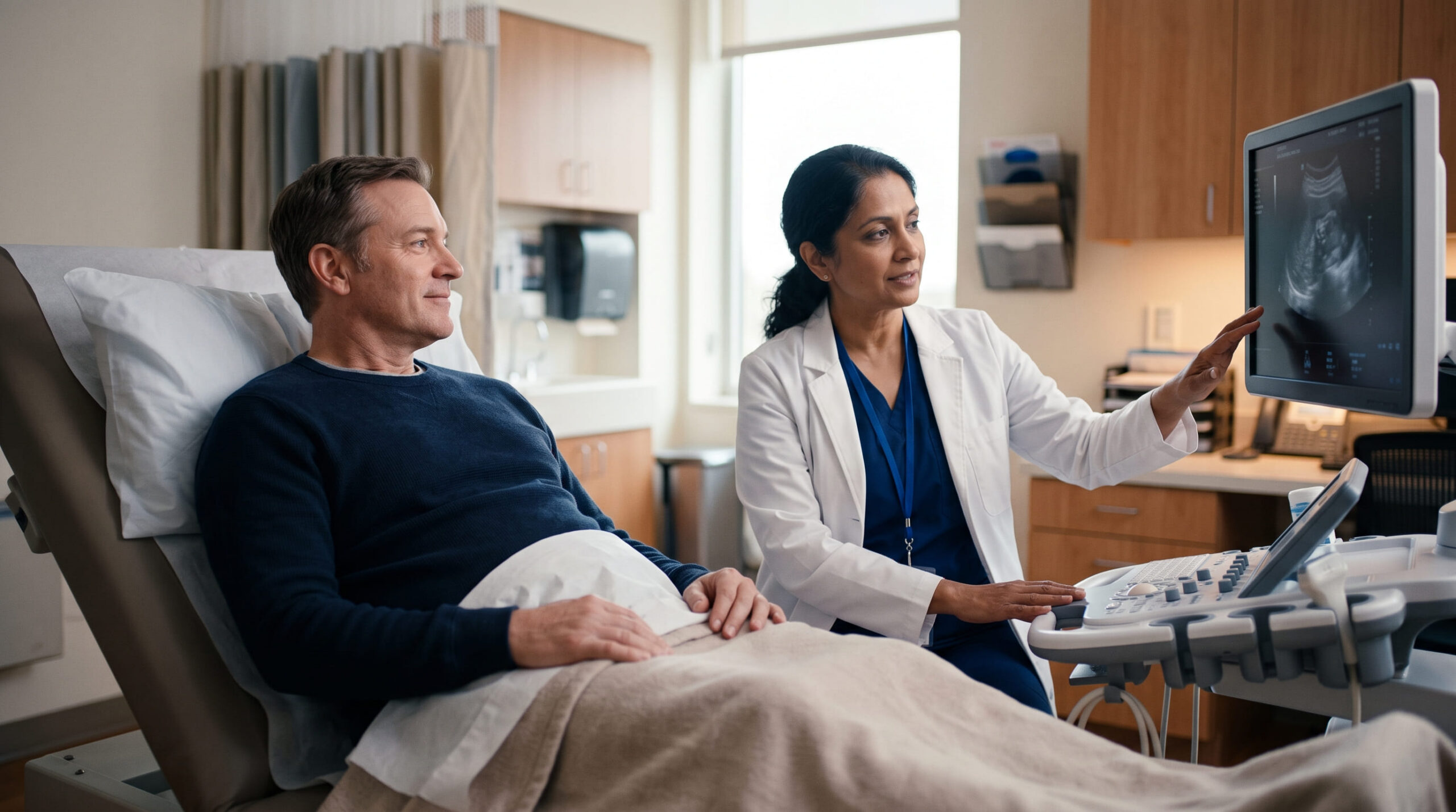
A kidney biopsy removes one or more tiny samples of kidney tissue, usually with a thin needle guided by ultrasound, so a pathologist can examine them under the microscope and tell doctors exactly what's damaging the kidney.

Briefly about this treatment
Indications
Persistent blood or protein in the urine, kidney function that's declining for no clear reason on blood and urine tests alone, a transplanted kidney that isn't working as well as expected, or an unusual mass found on a kidney scan.
Risks
Bleeding, pain at the biopsy site, and rarely infection or injury to a nearby structure. Risks are minimised by ultrasound guidance, careful patient selection and experienced teams.
Pre-procedure
Blood tests to check clotting and kidney function, a blood pressure check, pausing certain blood-thinning medicines on your doctor's instructions, and fasting for several hours beforehand.
Follow-up
Several hours of monitoring straight after the procedure, then a follow-up visit once the pathology report is ready (commonly one to two weeks) to go through what the tissue showed and plan next steps.
What is Kidney biopsy (renal biopsy)? An expert explains
Most kidney problems start with a clue on a lab test β blood or protein in the urine, or a creatinine level that’s climbing. Those tests tell your doctor that something is wrong, but not always what. A kidney biopsy answers the „what”: it gives a nephrologist a tissue-level view of inflammation, scarring, infection, or abnormal protein deposits, which is often the only way to reach a precise diagnosis and choose the right treatment.
During a standard percutaneous biopsy, a doctor β usually a nephrologist or an interventional radiologist β numbs a small area of your back with local anaesthetic, then uses ultrasound (sometimes CT) to guide a fine, hollow needle into the kidney and withdraw one or more thin cores of tissue, each about the size of a grain of rice. A renal pathologist then studies the sample under three types of microscopy β light, immunofluorescence and electron β to identify exactly which disease process is at work. The needle part of the procedure usually takes only a few minutes; the full visit, including prep and monitoring, is closer to half a day.
Signs it may be time for Kidney biopsy (renal biopsy)
Many people arrive at this page after a routine blood or urine test turned up something unexpected, not because they feel unwell. Watch for:
- Blood in your urine (visible, or picked up on a dipstick test) that doesnβt have an obvious cause
- Foamy or bubbly urine, which can signal high levels of protein (proteinuria)
- Swelling in your legs, ankles or around your eyes
- Unusual tiredness or fatigue that doesnβt have another explanation
- A creatinine level thatβs been rising, or an eGFR thatβs been falling, on repeat blood tests
On their own, these signs donβt mean you need a biopsy β most people with them are managed with monitoring, blood pressure control and medication first. A biopsy becomes relevant when those first steps donβt explain whatβs happening, or when the specific disease needs a tissue diagnosis to guide treatment.
These are the conditions linked to this treatment in the directory. Your clinician decides whether treatment is appropriate using your symptoms, examination and test results.
- Lupus Nephritis β Kidney inflammation from systemic lupus erythematosus.
- Vasculitis (ANCA-Associated) β Blood vessel inflammation damages kidneys.
- Cryoglobulinemic Glomerulonephritis β Immune complex deposition in glomeruli causing inflammation.
- C3 Glomerulopathy β Complement dysregulation causing glomerular injury.
- Henoch-SchΓΆnlein Purpura Nephritis β IgA vasculitis affecting kidneys.
Remember: These entries are not a diagnosis or a treatment recommendation. Discuss your symptoms and test results with a qualified clinician.
Β

When to consult a specialist
If a recent blood or urine test showed protein, blood, or a change in your kidney numbers, that’s a good reason to get it checked β not a reason to panic. A simple repeat blood test, urine test or ultrasound is often the sensible next step, and many causes turn out to be mild or manageable without ever needing a tissue sample. A kidney biopsy is something your nephrologist will only suggest if those first tests can’t explain what’s going on. Find a specialist to talk through your results and whether further testing, including a biopsy, makes sense for you.
How a session works
There are three main ways a kidney biopsy is performed, and your doctor will choose based on your anatomy and risk factors:
- Percutaneous biopsy (most common). A needle is passed through the skin over your lower back and guided into the kidney using real-time ultrasound (occasionally CT). Done under local anaesthetic, with light sedation offered in some cases.
- Transjugular biopsy. A thin catheter is threaded through a vein in the neck down into a kidney vein, and the tissue sample is taken from inside the blood vessel. This route is used when bleeding risk is higher than usual β for example, with certain clotting problems or severe obesity β because it avoids passing a needle through the skin and other tissue.
- Open or laparoscopic biopsy. A surgical option using a small incision, reserved for situations where a needle biopsy isn’t safe or possible, or when a larger piece of tissue is needed. This is done under general anaesthesia.
Benefits
A kidney biopsy is one of the few tests that shows doctors exactly what’s happening inside kidney tissue, not just what’s reflected in blood and urine numbers. That precision can:
- Confirm a diagnosis that other tests can’t reach on their own
- Reveal how active or advanced a kidney disease is, which shapes how urgently it needs treatment
- Guide the choice of treatment β for example, whether immunosuppressive medication is likely to help
- Help track whether a treatment is working, or whether a transplanted kidney is being rejected
For many patients, a biopsy is what turns an uncertain, worrying set of lab results into a clear plan they and their nephrologist can act on.
Risks & side effects
The treatment has risks and trade-offs to discuss with your care team. The information below comes from this treatmentβs data. Bleeding, pain at the biopsy site, and rarely infection or injury to a nearby structure. Risks are minimised by careful patient selection, ultrasound guidance and experienced teams.
- Blood in the urine. The most common side effect β light blood-staining is expected after the procedure and usually settles within 24 hours.
- Pain or soreness at the biopsy site. Usually mild; simple painkillers such as paracetamol are often enough.
- Bleeding that needs treatment. Uncommon; a small proportion of patients need a blood transfusion, and a smaller number need a follow-up procedure to stop bleeding that doesnβt settle on its own.
- Delayed bleeding. Rare, but it can appear up to about three weeks after the biopsy β worsening pain, blood in the urine, shortness of breath or light-headedness need urgent attention even weeks later.
- Infection. Uncommon, and usually treatable with antibiotics if it occurs.
- Injury to a nearby organ or blood vessel, loss of the biopsied kidney, or death. Extremely rare, but your doctor should discuss these possibilities as part of informed consent.
Who it may not suit: These are general cautions, not a personal verdict. A qualified clinician must assess your full medical picture before recommending or delaying treatment. A biopsy may be delayed or approached differently if you have an uncontrolled bleeding or clotting disorder, uncontrolled high blood pressure, only one working kidney, severe obesity or spinal deformity, an active urinary tract or kidney infection, or a kidney mass where imaging strongly suggests cancer.
Β

What happens during & after
- You check in, an IV line is placed, and your vital signs are checked; blood pressure medicine may be given if needed.
- You’re moved to the ultrasound or procedure suite and positioned β usually lying on your stomach, or on your side for a transplanted kidney.
- The skin is cleaned and numbed with local anaesthetic.
- Using ultrasound guidance, the doctor inserts the biopsy needle and collects one or more small tissue cores, often asking you to hold your breath for a few seconds during each pass to keep the kidney still.
- Firm pressure is applied to the site once enough tissue is collected.
- You’re taken to a recovery area and monitored lying flat for several hours (commonly 4β6, occasionally overnight) while your vital signs and urine are checked for bleeding.
- If everything looks stable, you’re discharged the same day with someone else driving you home; an overnight stay is sometimes recommended for higher-risk patients.
Who performs it: typically a nephrologist or an interventional radiologist performs the biopsy itself, working alongside a renal pathologist who examines the tissue afterward.
Possible complications
Most complications are limited to bleeding at or near the biopsy site, and are picked up during the hours of monitoring that follow the procedure. Less common ones include:
- Arteriovenous fistula. A small tear between a tiny artery and vein inside the kidney; it usually heals on its own without treatment and is occasionally found incidentally on a later scan.
- Bleeding needing intervention. A blood transfusion, or a follow-up procedure such as embolisation, is uncommon but sometimes needed to stop bleeding that doesn’t settle by itself.
- Infection. Uncommon, and generally responds well to antibiotics.
- Surgery. Rarely, significant bleeding needs a surgical repair rather than a minimally invasive fix.
Serious complications requiring transfusion, a procedure, or surgery are uncommon, which is why patients are monitored closely for several hours before being sent home.
New or severe symptoms require prompt medical advice. Do not wait for a routine appointment if you feel seriously unwell.
Recovery
Right after the procedure: You’ll rest lying flat for several hours in a recovery area while nurses check your blood pressure, pulse and biopsy site, and look at your urine for signs of bleeding. This is the highest-risk window for bleeding, which is why most patients are watched closely before being allowed home.
At home β the first one to two weeks:
- Day 1: Rest at home. Avoid driving, operating machinery, or making important decisions if you were sedated.
- First 24β48 hours: Take it easy β light walking is fine, but avoid bending, straining, or heavy lifting.
- First 1β2 weeks: Avoid strenuous exercise, contact sports, heavy lifting, and any blood-thinning medicines or supplements unless your doctor has said it’s safe to restart them. Drink plenty of water, and use simple painkillers such as paracetamol for soreness at the site as needed.
Your doctor will tell you when it’s safe to return to normal activity, work and exercise β this varies by individual and by biopsy type.
Warning signs β contact your doctor or go to the ER if you notice heavy, dark or persistent blood in your urine or blood clots, difficulty or inability to pass urine, worsening pain at the biopsy site or in your side or back, redness, swelling, warmth or drainage at the site, a fever or chills, or feeling faint, dizzy or short of breath. Because serious bleeding can occasionally appear as late as about three weeks after the biopsy, keep an eye out for these signs even after you feel fully recovered.
Getting your results: Some initial findings may be available within a few days, but a full pathology report β including specialised immunofluorescence and electron microscopy β commonly takes one to two weeks. Your nephrologist will usually go over the results with you at a follow-up appointment and explain what they mean for your treatment plan.
This is general guidance. Follow the specific instructions given by your own care team, which may differ based on your biopsy type and health history.

Specialists for Kidney biopsy (renal biopsy)
Specialists linked to this treatment in the KidneyGlobal directory. Review credentials and confirm availability directly before booking.
Verified specialists


Clinics offering Kidney biopsy (renal biopsy)
Verified kidney centres linked to this treatment in the KidneyGlobal directory.


Most Common Questions
Is a kidney biopsy painful?
Most people feel pressure rather than sharp pain, because the skin and the area around the kidney are numbed with local anaesthetic first. Some soreness at the site afterward is common and usually manageable with simple painkillers such as paracetamol.
How long does a kidney biopsy take?
The needle procedure itself is typically 30β60 minutes, including positioning and ultrasound guidance, though the actual tissue sampling takes only a few minutes. Including check-in, prep and recovery monitoring, plan for most of a day.
Will I be awake during the procedure?
Usually, yes. Most kidney biopsies are done under local anaesthetic, sometimes with a light sedative to help you relax; general anaesthesia is reserved for less common approaches, such as an open surgical biopsy.
How long until I get my results?
Initial findings are sometimes available within a few days, but a complete report β which needs specialised staining and electron microscopy β commonly takes one to two weeks. Your nephrologist will usually go through the results with you at a follow-up visit.
Can a kidney biopsy be done on a transplanted kidney?
Yes. A transplant kidney biopsy is one of the most reliable ways to check whether a transplanted kidney is being rejected or has another problem, and is often used either as a scheduled check or when blood tests suggest something has changed.
Is a kidney biopsy safe?
It's considered a safe, well-established procedure, though like any procedure it carries some risk, mainly bleeding. See the Risks & Side Effects section for a full breakdown, and always discuss your personal risk factors with your doctor before the procedure.
Sources
- Kidney BiopsyNational Institute of Diabetes and Digestive and Kidney Diseases (NIDDK), NIH
- Kidney biopsy: OverviewMayo Clinic
- Kidney BiopsyCleveland Clinic
- Kidney BiopsyNational Kidney Foundation
- Kidney BiopsyNational Kidney Federation (UK)