

Academic Medical Center
Comprehensive Nephrology & Transplant
Joint CommissionMagnet RecognitionUNOS Transplant Center
✓ 24/7 ER✓ ICU✓ Insurance
Cryoablation is a minimally invasive, image-guided procedure that freezes and destroys a small kidney tumor without removing the kidney, letting many people avoid open surgery and go home the same day or after one night.
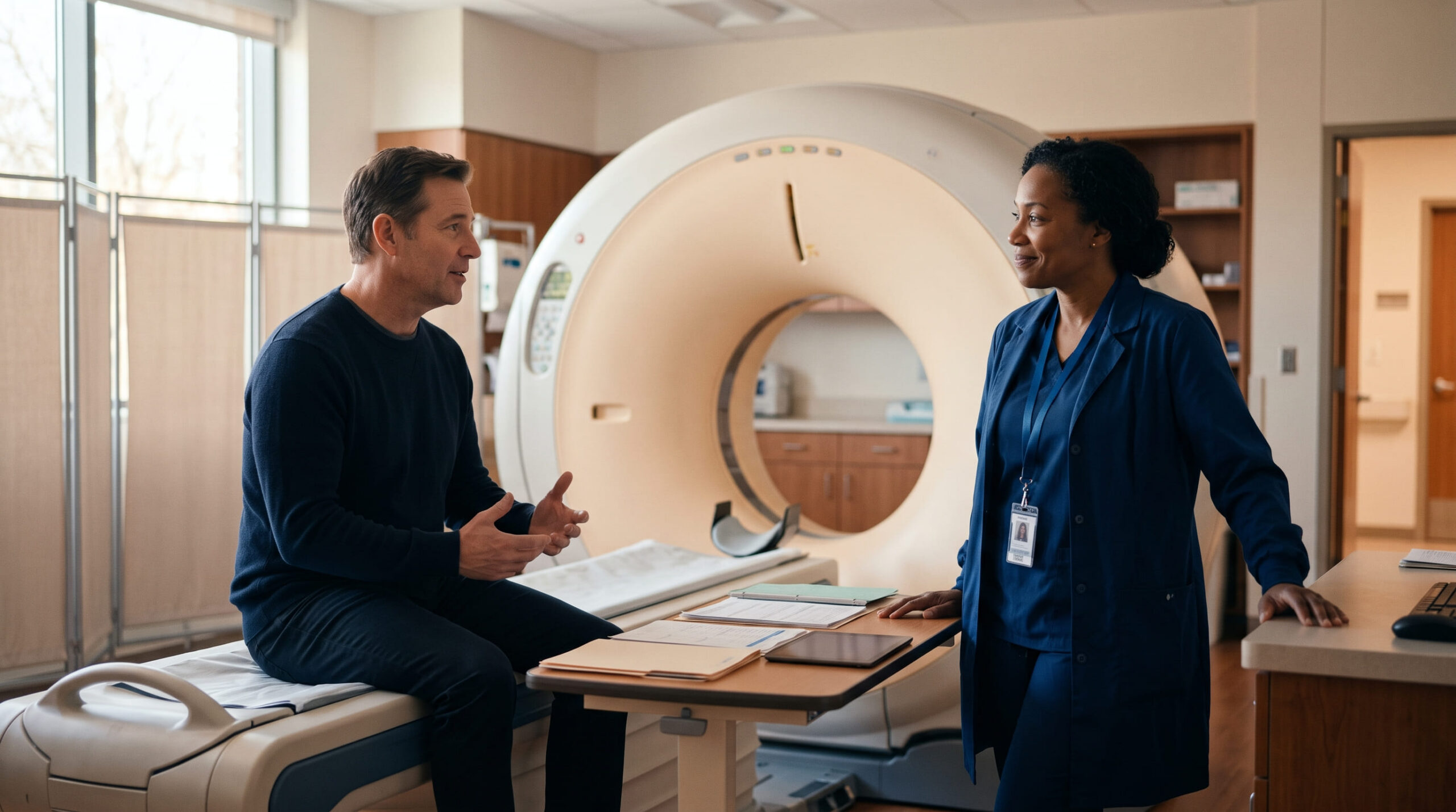
A small kidney tumor (generally under 4 cm), a wish to avoid the anesthesia and recovery of open surgery, a single or poorly functioning kidney, multiple tumors from an inherited condition, higher surgical risk from age or other health conditions, or a tumor that has come back after previous surgery or ablation.
Bleeding or bruising around the probe site, infection, and rare injury to a nearby organ or the urine-collecting system. Risks are minimised by careful patient selection, imaging guidance and experienced interventional radiology teams.
Standard blood work, a planning CT or MRI scan (often with a needle biopsy of the tumor), an anaesthesia consultation, and fasting per your center's protocol.
Imaging (CT or MRI) at intervals over the first few years to confirm the tumor stays destroyed, with kidney function checked by blood test at the same visits.
An interventional radiologist — a doctor who treats disease using imaging instead of open surgery — guides one or more hollow probes through the skin and into the tumor using CT, MRI or ultrasound. Gas pumped through the probes drops the tumor’s temperature to roughly -20°C to -40°C, forming a visible „ice ball” that kills the tumor cells. The tissue is allowed to thaw, then frozen again — usually two freeze–thaw cycles — which is what destroys the tumor for good. The kidney tissue around the ice ball is spared.
You don’t choose cryoablation because of symptoms. Most small kidney tumors cause none at all and are found by chance on a scan done for another reason. Still, a few things are worth mentioning to your doctor:
These symptoms have many possible causes, but imaging can rule a kidney tumor in or out.
These are the conditions linked to this treatment in the directory. Your clinician decides whether cryoablation is appropriate using your symptoms, examination, imaging and test results.
Remember: These entries are not a diagnosis or a treatment recommendation. Discuss your symptoms and test results with a qualified clinician.

Finding a small kidney mass on a scan is unsettling, but most are caught early and there are good options, including this one. A simple blood and urine check, alongside the imaging you already have, gives a specialist most of what they need for a first opinion — there’s no need to wait for things to feel worse. If your doctor has flagged a renal mass, or you’re managing a hereditary condition linked to kidney tumors, find a specialist on KidneyGlobal to talk through whether cryoablation, surveillance or surgery fits your situation.
Cryoablation is usually done as a single outpatient or overnight-stay session, typically lasting 1–2 hours depending on tumor size and number of probes needed.
Planning scan and biopsy. A contrast CT or MRI maps the tumor’s size and position. Many centers take a needle biopsy of the tumor — either beforehand or at the start of the same session — to confirm what type of tissue is being treated.
Anesthesia and positioning. Most procedures use general anesthesia or deep sedation; some centers use local anesthesia with light sedation. You’re positioned (often lying on your front or side) so the radiologist has a clear, safe path to the kidney.
Probe placement. Using real-time CT, MRI or ultrasound guidance, the radiologist inserts one or more thin cryoprobes through the skin directly into the tumor. The number of probes depends on tumor size — larger tumors need more probes to freeze completely.
Protecting nearby structures. If the tumor sits close to the bowel, ureter or other organs, the team may inject saline or air between structures (hydro- or pneumo-dissection) to create a safety margin before freezing starts.
Freeze–thaw cycles. Pressurized argon gas cools the probe tips, forming an „ice ball” that engulfs the tumor plus a small margin of surrounding tissue. The team watches the ice ball grow on live imaging. The tissue is then allowed to thaw (often helped by pumping helium gas through the probes), and the freeze is usually repeated once more to make sure the tumor is fully treated.
Probe removal and closure. Once imaging confirms the ice ball fully covers the tumor with an adequate margin, the probes are withdrawn and the small skin punctures are closed with a bandage — no stitches are usually needed.
Recovery room monitoring. You’re watched for a few hours for bleeding, pain or changes in vital signs before either going home the same day or staying one night for observation, depending on your center’s protocol and your health status.
Compared with surgery, cryoablation offers a gentler path for many people with a small kidney tumor. Guideline bodies including the AUA, NCCN and NICE recognize thermal ablation as a reasonable option for small kidney tumors — particularly for people who are frail, have a solitary kidney, reduced kidney function, or a genetic condition causing multiple tumors — while partial nephrectomy generally remains the standard for fit surgical candidates.
The treatment has risks and trade-offs to discuss with your care team. The information below comes from this treatment’s own data: bleeding or bruising around the probe site, infection, and rare injury to a nearby structure. Risks are minimised by careful patient selection and experienced teams. Most complications are minor and resolve on their own; serious complications are uncommon, and your own risk depends on tumor size, location and your overall health.
Who it may not suit: these are general cautions, not a personal verdict. A qualified clinician must assess your full medical picture before recommending or delaying treatment.

During the procedure you’re asleep or sedated, so you won’t feel the freezing. Afterward, you wake in a recovery area where staff check your vital signs, pain level, and the puncture sites for bleeding. Many patients go home the same day; some stay one night for observation, especially after treatment of a larger tumor or if you live far from the hospital.
The first week. Expect mild to moderate soreness at the puncture sites and possibly some fatigue for a few days. Most people return to light daily activity within a few days and to normal activity, including work, within about a week — much faster than after open or laparoscopic surgery. Your team will give you specific instructions on:
Most people recover without incident. Complications that can occur in the days to weeks afterward include:
Call your care team (or seek urgent care) if you develop fever, spreading redness or drainage at the puncture site, heavy or worsening bleeding, or severe pain that isn’t controlled by your prescribed medication.
New or severe symptoms require prompt medical advice. Do not wait for a routine appointment if you feel seriously unwell.
Recovery from cryoablation is measured in days, not weeks. Most people return to light daily activity within a few days and to normal activity, including work, within about a week.
Because the tumor is destroyed in place rather than removed, follow-up CT or MRI scans are how your team confirms the treatment worked and stays working, rather than a single pathology report. Your clinician will set a personal schedule — typically imaging at intervals over the first few years and then periodically afterward, rather than a one-size-fits-all timetable. Kidney function (via blood tests) is usually rechecked at the same visits.
Verified kidney centres linked to this treatment in the KidneyGlobal directory.



No. Nothing is removed. The tumor is destroyed in place, and the dead tissue is gradually reabsorbed or remains as scar tissue, monitored on future scans.
For small, localized tumors, published series report high local tumor-control rates, but ablation is a local treatment, not a guarantee — your care team will set a personal follow-up imaging schedule to confirm the tumor stays destroyed. This page is educational and isn't a substitute for advice from your own clinician about your own scans.
Most people are under general anesthesia or deep sedation during the procedure itself, so they feel nothing at the time. Mild soreness at the probe site for a few days afterward is common.
Both are image-guided needle treatments for small kidney tumors. Cryoablation freezes tissue; RFA heats it. Guideline bodies consider both reasonable options for suitable tumors, and the choice often comes down to tumor location and your center's experience.
Usually an interventional radiologist performs the procedure itself; a urologist or urologic oncologist typically refers you, confirms the diagnosis, and follows you afterward.
Yes, and it's reasonable to ask for one. Because ablation and surgery both have a role depending on tumor size, location and your health, a second opinion from a urologic oncologist is a normal part of the decision, not a sign something is wrong.